Sunday, April 18, 2010

Osgood-Schlatter Disease


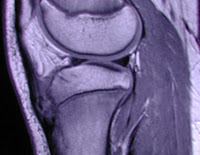
Osgood-Schlatter disease is a condition where the patellar tendon of the knee has been pulled from the tibia. This disease usually happens in males during adolescence. Most often than not, this condition occurs from a repetitive stress placed on the tendon, such as running or jumping. However, this disease can also occur from rapid growth.

Symptoms include pain, especially when exercising, limping, sensitive to touch, swelling, and sometimes avulsion fractures of the tibia. Most of the time this disease will heal itself, but sometimes further treatment is needed. Rest, ice, elevation, anti-inflammatory medications, stretching the quadriceps muscles slowly, stabilizing the knee by a brace, and surgery to re-attach the tendon are possible treatments.


References:
http://www.mskcases.com/images/article/1/113-745.JPG


Sunday, March 21, 2010

Separated shoulders


A separation of the shoulder is most often caused from contact sports. A schoulder separation is an injury involving the acromion process of the clavicle and the coracoid process of the scapula, also called the acromioclavicular joint (AC). Damage is dependendant on the extensiveness of the injury. There are three ligaments involved in this area. One is the acromioclavilar ligament (AC), one is the coracoiclavicular ligament (CC), and the last is the coracoacromial ligament.


X-rays are commonly performed first; then, if needed, an MRI scan is done to see the damage of the displacement. Shoulder separations are graded from 1 to 6. One is having the least damage and six is having the most damage. A rupture of both the AC ligament and the CC ligament is known as a grade 6 separation. Most often surgery will have to be done to fix this problem.


Some symptoms include: obvious dislocation, pain, swelling, and not being able to move the joint.


Treatments are generally to put the bones back into place, but surgery can be done if the separation is bad enough. A specific type of surgery is called an arthroscopic reconstruction. In this surgery, torn ligaments can be grafted to provide excellent results for the patient. Anti-inflammatory medication can be taken to decrease swelling. Splinting the shoulder with a shoulder immobilizer and letting the shoulder heal is another possible treatment. Of course the most obvious treatments are rest and ice. Sometimes patients may have complications with certain types of treatments, which may include: clavicle fracture, infection, scar, pain, and deltoid or trapezius muscle detachment. They may have to have further care and possibly physical therapy.


The image I have uploaded is an MRI T2 weighted fat saturation coronal image. The ligament tear is noted, which causes an AC separation. It is also noted that there is a highlighted area, this is inflammation in the joint space that should not be there!


References:

http://www.sportsinjuryclinic.net/

Image is from:radiology.med.miami.edu/x69.xml

Friday, February 12, 2010

Ovarian cysts


In the year 2000, my aunt and my mother convinced my grandmother to go to the doctor to have a check up. They had been noticing that my grandmother was looking sick and gaining a lot of weight in a short period of time. She only complained of having some minor back pain, but after not going to a doctor for about 30 years, one would start to get used to pain. After being examined by the doctor, she had a CT scan that showed a huge ovarian cyst. The doctor soon scheduled her an appointment with a general surgeon to remove the cyst. They took pictures before and during the surgery. I have uploaded actual pictures of my grandmothers cyst! Recently when I was talking to my grandmother, she said her cyst weighed 29.5 lbs! The doctors that performed the surgery said they had only seen one that was larger, which was 30 lbs.

Ovarian cyst are composed of fluid such as blood, mucous, etc. found inside the ovaries. There are two types of ovarian cysts, one being a follicular cyst, which is basically enlarged follicles, and the other known as a corpus luteum cyst, which is basically a hemorrhage of the corpus luteum (the lining within the ovary). Ovarian cysts are more common in females around childbearing age, but is definitely not uncommon to find them in older women.


Lower pelvic pain, back pain, abnormal bleeding, fullness or a heavy feeling in pelvic area, and painful intercourse are just some of the main symptoms of having ovarian cysts. There can be one or multiple. Misdiagnosis can often occur because doctors mistake it for menstrual cramps. If a cyst ruptures it can cause extreme pain and


Ultrasound is the best diagnostic imaging tool to use, but cysts can also be seen in CT or MRI exams.
I have uploaded MRI images. The top image is an example of an ovarian cyst in a T1-weighted image, the bottom is a T2-weighted image (shows a greater signal intensity).

Surgery is done in extreme cases, but the pain can be masked with a narcotic or even ibuprofen. Birth control pills can be given to the patient to help prevent the formation of new cyst.


References:


Eisenberg, Ronald & Johnson, Nancy (2003) Comprehensive Radiographic

http://www.glowm.com

A special thanks is given to my grandmother for letting me use these images.

Saturday, February 6, 2010

Appendicitis

When I was 12 years old, it seemed like I was sick all the time. My stomach/abdomen would hurt off and on for months. My primary care physician ordered an upper GI and small bowel follow through, but there were no abnormal findings. Then, one morning I woke up with excruciating abdominal pain and could not get out of the fetal position. I was running a temperature, nausea, vomiting, and it burned so badly to urinate. My mother just thought that I had the flu because flu season was going around, but the reality was, I had chronic appendicitis for a few months until the day my appendix ruptured. I developed gangrene and had it for three days! When I got out of my 4-hr surgery to remove my appendix and some of my bowel, the doctors said I was less than 4 hours from dying! I am so lucky to still be alive today. If it wasn't for the surgeons that day, I would've been dead!
Appendicitis is an inflammation of the appendix, which is found at the junction of the small and large intestines and is located on the cecum. It can be either acute (sudden onset) or chronic (happens over a period of time). Genetic predisposition plays a small role in developing appendicitis, especially if the person has a mother, father, or sibling that has had appendicitis. Anatomy position of their appendix plays another role.
Appendicitis occurs because of food particles or other types of particles gets lodged in the appendix and causes an obstruction that eventually leads to infection. Also lymph nodes can enlarge and cause an obstruction. If the obstruction is left untreated it can cause peritonitis, which is what I experienced. Surgery to remove the appendix is always performed. In my case they had to completely open me up to remove my appendix, but in a lot of cases where the appendix has not been ruptured, they can remove it laproscopically.
Symptoms include: nausea, vomiting, pain originating in periumbilical area toward the right lower quadrant or as it is commonly referred as "McBurney's point", low-grade fever, elevated white blood cell count, etc.
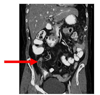
CT scans are probably one of the better modalities for looking at the appendix. Coronal re formats in CT are very helpful in reading these exams. Radiologist measure the appendix by its' width and if it measures over 6 mm, it is appendicitis. It can sometimes be tricky to determine whether or not a patient has appendicitis because the position of the appendix is located in differently in every patient. Sometimes the appendix can be so large, it looks like a continuation of the large bowel and sometimes it can lie directly on the psoas muscle and be very long. Radiologist can sometimes mistake appendicitis for a specific lymph node enlargement called mesenteric lymphadenitis, because its appearance is similar to appendicitis.

I have uploaded an example of a CT coronal image. The red arrow is pointing to the enlarged appendix.

References:

Dr. Mitchell, MD
www.emedmag.com
http://radiology.rsna.org

Saturday, January 23, 2010

Aortic Dissection


A very close friend of mine was complaining of sudden onset tooth pain then quickly changed to excruciating chest pain. His family rushed him to the nearest emergency room, where he had an EKG (which came back normal), so a chest CT scan performed with contrast to rule out an aneurysm. The end result was actually a dissection of the ascending aorta. ER staff immediately sent for a helicopter to take him to a very well known hospital in Indianapolis. At the hospital, the doctors were able to fix his dissection through surgery by placing a nylon patch over the dissection. He was in intensive care for 4-5 days, but was over medicated with coumadin, so they had to keep him an extra few days to make sure he was fine. It is amazing to me that he is still living today and that he walked out with only one medication that he takes, which is toprol.

The aorta is the largest artery in the body and arises from the heart and it is made of several layers. A dissection is caused from a tear of the layers in the wall of the aorta. Anyone can have a tear in the wall of the aorta, but it is definately more common in men between the ages of 60-70. It can also occur in post traumatic situations. High blood pressure, hyperlipidemia, weakening of arterial walls, etc are all examples of risk factors for having a dissection.

There are two types of aortic dissection. The first, type A, is where the tear is on the ascending aorta, the second, type B, is where the tear is on the descending side of the aorta. Type A is difinately more crucial because there is a lot more blood flow and pressure at this point.

Surgery can be done to correct the dissection by placing a wire mesh material through the aorta. Medication can also be given to the patient to help a chronic dissection.

References:

Mayoclinic.com
Dr. Mary C Mancini, MD, PhD at webMD.com
www.imagingpathways.health.wa.gov

Wednesday, January 13, 2010

Emphysema


Emphysema is a chronic obstructive pulmonary disease that is progressive and irreversible. When a patient develops emphysema, the air literally becomes trapped within the lungs because the alveoli (where CO2 is exchanged for O2) starts to collapse. Patients also develop a "barrel chest", which can be seen on a general PA chest x-ray. This "barrel chest" is formed by the rounding of the costophrenic angles.

Some other symptoms may include very short and rapid breaths, shortness of breath, chest pain/tightness, coughing, and fatigue.

My grandfather was a wonderful man, but he had a bad habit. He smoked for years and years. He eventually developed emphysema and his life was shortened after catching pneumonia for a second time within one year. He was in and out of intensive care that year and for his last 5 years he had to carry an oxygen tank with him every where he went. He told my grandmother to "pull the plug" if he ever had to go back under the ventilator. He definitely did not want to live his life like that, and to be honest, who would?

In April of 2004, I went to visit my grandfather in the hospital. I was actually the last grandchild he spoke with before he went into a coma. He told me he was so proud of my sister and I for going to college, being great, honest people, and for not ever smoking. I wish that I could have done more for him and I really wish I would have spent more time with him before he passed away. He was in a lot of pain there at the end! His kidneys failed after he went into a coma, so my grandmother decided it was best to take him off the ventilator. Smoking is such an awful thing to do to your body. I seriously wish everyone would understand that and quit!

References: www.mayoclinic.com, www.ctsnet.org, www.mevis-research.de
Sunday, December 13, 2009

Low back pain


One of my closest friends has had several back issues throughout her life. She used to be a competitive figure skater. For starters she has a history of a healed compound fracture of L-3 that was just diagnosed, which has caused some wedging of the vertebra. When she was eleven, she was diagnosed with scoliosis from L-3 through L-5 due to repetitive trauma to her growth plate of the left hip from her skating. The fifth lumbar vertebra is taking the same form of the sacrum causing what is called L-5 sacralization. (The image on the right shows an example of this.) And if matters couldn't get worse, she also has spina bifida occulta, which is where the spinous process on the inside of the vertebra is not closed properly. My friend is actually very lucky, because in some cases, spina bifida occulta can lead to huge deformities of the legs, feet (an example is clubfoot), and the spine making the patient unable to walk or walk without the use of a walking device. Throughout her whole life, she has had numerous MRI scans to monitor her condition.

What I found most interesting when talking with her, is that patients with spina bifida occulta can have several conditions that result from having this disorder. Some of these conditions are: UTI's, skin infections, and skin allergies.

She has tried several means of management to minimize pain and mazimize daily function. Initially, after the growth plate injury, she tried physical therapy with ultrasound and rest. She also was fitted for orthotic inserts to build her left side up to match the right so she doesn't walk crooked. Then she tried occupational therapy or "spinal re-training". She even had a cortisone injection under fluoroscopy, but unfortunately, it didn't help. Today, she uses a combination of chiropractic care, massage therapy, and a TENS unit (trans-cutaneous electro-neuromuscular stimulation). All of these treatments have helped tremendously with the pain and spasms, but she occasionally has to resort to using oral pain medication. Thankfully my friend has never had to have surgery, but some patients with these deformities have to undergo the knife, especially with severe cases of spina bifida occulta or growth plate trauma.

References:

www.ajronline.org