When I was 12 years old, it seemed like I was sick all the time. My stomach/abdomen would hurt off and on for months. My primary care physician ordered an upper GI and small bowel follow through, but there were no abnormal findings. Then, one morning I woke up with excruciating abdominal pain and could not get out of the fetal position. I was running a temperature, nausea, vomiting, and it burned so badly to urinate. My mother just thought that I had the flu because flu season was going around, but the reality was, I had chronic appendicitis for a few months until the day my appendix ruptured. I developed gangrene and had it for three days! When I got out of my 4-hr surgery to remove my appendix and some of my bowel, the doctors said I was less than 4 hours from dying! I am so lucky to still be alive today. If it wasn't for the surgeons that day, I would've been dead!
Appendicitis is an inflammation of the appendix, which is found at the junction of the small and large intestines and is located on the cecum. It can be either acute (sudden onset) or chronic (happens over a period of time). Genetic predisposition plays a small role in developing appendicitis, especially if the person has a mother, father, or sibling that has had appendicitis. Anatomy position of their appendix plays another role.
Appendicitis occurs because of food particles or other types of particles gets lodged in the appendix and causes an obstruction that eventually leads to infection. Also lymph nodes can enlarge and cause an obstruction. If the obstruction is left untreated it can cause peritonitis, which is what I experienced. Surgery to remove the appendix is always performed. In my case they had to completely open me up to remove my appendix, but in a lot of cases where the appendix has not been ruptured, they can remove it laproscopically.
Symptoms include: nausea, vomiting, pain originating in periumbilical area toward the right lower quadrant or as it is commonly referred as "McBurney's point", low-grade fever, elevated white blood cell count, etc.
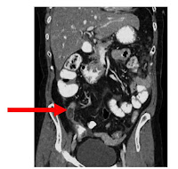
CT scans are probably one of the better modalities for looking at the appendix. Coronal re formats in CT are very helpful in reading these exams. Radiologist measure the appendix by its' width and if it measures over 6 mm, it is appendicitis. It can sometimes be tricky to determine whether or not a patient has appendicitis because the position of the appendix is located in differently in every patient. Sometimes the appendix can be so large, it looks like a continuation of the large bowel and sometimes it can lie directly on the psoas muscle and be very long. Radiologist can sometimes mistake appendicitis for a specific lymph node enlargement called mesenteric lymphadenitis, because its appearance is similar to appendicitis.
I have uploaded an example of a CT coronal image. The red arrow is pointing to the enlarged appendix.
References:
Dr. Mitchell, MD
www.emedmag.com
http://radiology.rsna.org


No comments:
Post a Comment